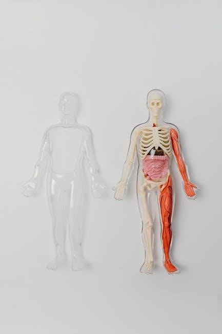
Anatomy and Physiology Coloring Workbook serves as an excellent study guide‚ simplifying complex concepts through engaging visuals and interactive learning experiences for students.
Combining a wide variety of engaging coloring activities‚ these workbooks enhance understanding of the human body’s intricate structures and functions.
These resources feature detailed illustrations‚ aiding in memorization and providing a unique approach to mastering anatomy and physiology coursework effectively.
What is an Anatomy and Physiology Coloring Workbook?
An Anatomy and Physiology Coloring Workbook is a unique study tool designed to reinforce learning through visual and kinesthetic engagement. Unlike traditional textbooks‚ these workbooks present anatomical structures and physiological processes as line drawings‚ requiring students to actively color and label.
This process isn’t merely artistic; it significantly enhances memory retention and comprehension. By physically interacting with the material‚ students move beyond rote memorization and develop a deeper understanding of the body’s complexities. These workbooks often include detailed illustrations by renowned medical illustrators like Drs. Frank H. Netter and Carlos Machado‚ ensuring accuracy and clarity.
They typically cover a broad range of body systems‚ from the skeletal and muscular systems to the nervous and cardiovascular systems‚ offering a complete study guide for anatomy and physiology courses. The workbooks are designed for both introductory and advanced levels‚ catering to diverse learning needs.
Benefits of Using a Coloring Workbook
Utilizing an Anatomy and Physiology Coloring Workbook offers numerous pedagogical advantages. The act of coloring actively engages the brain‚ promoting deeper learning and improved retention compared to passive reading. This kinesthetic learning style caters to diverse learners‚ enhancing comprehension of complex anatomical structures and physiological processes.
Color-coding allows for the organization of information‚ making it easier to differentiate between various components and systems. Workbooks simplify the study of anatomy and physiology‚ providing a less intimidating approach to challenging subject matter. They serve as a complete study guide‚ supplementing lectures and textbooks effectively.
Furthermore‚ coloring reinforces memory through visual association‚ aiding in recall during exams. These workbooks are valuable for students in paramedic‚ EMT‚ and other healthcare programs‚ offering a practical and engaging learning experience.
Target Audience: Students and Professionals
Anatomy and Physiology Coloring Workbooks are designed for a broad audience‚ primarily targeting students enrolled in healthcare-related courses. This includes undergraduate students in biology‚ nursing‚ pre-med‚ and allied health programs who require a complete study guide to supplement their learning.
However‚ the benefits extend beyond the academic realm. Professionals such as paramedics‚ EMTs‚ and other healthcare practitioners can utilize these workbooks for continuing education and refresher courses. The detailed illustrations and interactive format aid in reinforcing anatomical knowledge and improving diagnostic skills.
Even individuals with a general interest in the human body can benefit from these resources‚ providing a visually engaging and accessible way to learn about anatomy and physiology. They cater to various learning styles and levels of expertise.
The Human Body Systems: A Coloring Journey
Coloring through each system – skeletal‚ muscular‚ nervous‚ cardiovascular‚ and more – provides a detailed‚ visual exploration of anatomy and physiology.
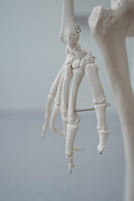
Skeletal System Coloring
Coloring the skeletal system offers a foundational understanding of bone structures‚ including long bones‚ short bones‚ flat bones‚ and irregular bones‚ alongside their specific features.
Detailed illustrations within anatomy and physiology coloring workbooks showcase various bones – femur‚ tibia‚ humerus‚ skull – allowing students to visualize their shapes and articulations.
This process reinforces knowledge of bone markings like processes‚ foramina‚ and fossae‚ crucial for understanding muscle attachments and nerve pathways.
Students can differentiate between axial and appendicular skeletons‚ learning the functions of each skeletal component in supporting the body and enabling movement.
Color-coding different bone regions aids in memorization and comprehension of the skeletal system’s complex anatomy‚ enhancing overall learning retention.
Muscular System Coloring
Coloring the muscular system provides a detailed exploration of muscle types – skeletal‚ smooth‚ and cardiac – and their unique structural characteristics.
Anatomy and physiology coloring workbooks present illustrations of major muscles like biceps brachii‚ quadriceps femoris‚ and gastrocnemius‚ aiding in visualization of their location and attachments.
Students learn to identify muscle origins‚ insertions‚ and actions‚ understanding how muscles work in antagonistic pairs to produce movement.
The process reinforces knowledge of muscle fiber arrangements – parallel‚ pennate – and their impact on muscle strength and range of motion.
Color-coding different muscle groups enhances memorization and comprehension of the muscular system’s complex anatomy‚ improving learning outcomes significantly.
Nervous System Coloring
Coloring the nervous system offers a detailed study of its intricate components‚ including the brain‚ spinal cord‚ and peripheral nerves‚ enhancing anatomical understanding.
Anatomy and Physiology Coloring Workbooks illustrate the central and peripheral nervous systems‚ aiding in visualizing neuronal structures like neurons and glial cells.
Students learn to differentiate between sensory‚ motor‚ and interneurons‚ understanding their roles in transmitting signals throughout the body effectively.
The process reinforces knowledge of brain regions – cerebrum‚ cerebellum‚ brainstem – and their associated functions‚ improving comprehension of neurological processes.
Color-coding different nerve pathways and brain areas aids memorization and facilitates a deeper understanding of the nervous system’s complex organization.
Central Nervous System
Coloring the central nervous system (CNS) – brain and spinal cord – provides a focused study of its structures and functional divisions‚ enhancing anatomical recall.
Anatomy and Physiology Coloring Workbooks detail brain regions like the cerebrum‚ cerebellum‚ and brainstem‚ illustrating their unique anatomical features.
Students visualize the layers of the brain‚ including the cerebral cortex and underlying structures‚ understanding their roles in higher-level cognitive functions.
The spinal cord’s anatomy‚ including gray and white matter‚ is clarified through coloring‚ aiding in understanding nerve pathways and reflex arcs.
Color-coding different brain lobes and spinal cord segments reinforces memorization and improves comprehension of the CNS’s complex organization and function.
Peripheral Nervous System
Coloring the peripheral nervous system (PNS) – nerves and ganglia outside the brain and spinal cord – clarifies its intricate network and functional components.
Anatomy and Physiology Coloring Workbooks illustrate cranial and spinal nerves‚ detailing their origins‚ pathways‚ and target destinations within the body.
Students visualize the somatic and autonomic nervous systems‚ differentiating between voluntary and involuntary control mechanisms through distinct color-coding.
The sympathetic and parasympathetic divisions of the autonomic nervous system are highlighted‚ demonstrating their opposing roles in regulating bodily functions.
Coloring sensory and motor neurons aids in understanding signal transmission and the PNS’s role in connecting the CNS to the body’s periphery effectively.
Cardiovascular System Coloring
Coloring the cardiovascular system‚ encompassing the heart‚ blood vessels‚ and blood‚ provides a visual understanding of circulatory pathways and functions.
Anatomy and Physiology Coloring Workbooks detail the heart’s chambers‚ valves‚ and major vessels‚ clarifying blood flow through the pulmonary and systemic circuits.
Students differentiate arteries‚ veins‚ and capillaries through color-coding‚ visualizing their structural differences and roles in oxygenated and deoxygenated blood transport.
The workbook illustrates the coronary arteries‚ highlighting their importance in supplying the heart muscle with oxygen and nutrients‚ preventing potential ischemia.
Coloring the lymphatic system alongside the cardiovascular system demonstrates its crucial role in fluid balance‚ immunity‚ and lipid absorption effectively.
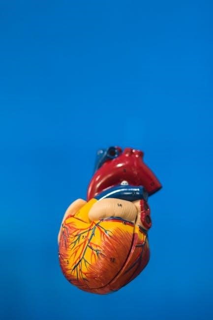
Heart Anatomy
Coloring the heart’s anatomy within Anatomy and Physiology Coloring Workbooks allows for detailed visualization of its four chambers – the right and left atria and ventricles.
Students identify and shade the atrioventricular valves (tricuspid and mitral) and semilunar valves (pulmonary and aortic)‚ understanding their function in unidirectional blood flow.
The workbook emphasizes the cardiac muscle (myocardium)‚ pericardium‚ and the layers of the heart wall‚ aiding in comprehension of its structural components.
Color-coding the coronary arteries and veins illustrates the heart’s own blood supply‚ crucial for understanding potential ischemic events and cardiac function.
Detailed illustrations showcase the sinoatrial (SA) node‚ atrioventricular (AV) node‚ and Purkinje fibers‚ clarifying the heart’s intrinsic conduction system effectively.
Blood Vessels
Coloring exercises in Anatomy and Physiology Coloring Workbooks focus on differentiating arteries‚ veins‚ and capillaries based on their structural characteristics.
Students visually distinguish between the thick-walled arteries‚ with their elastic layers‚ and the thinner-walled veins‚ often featuring valves to prevent backflow.
The workbooks illustrate the three layers of blood vessel walls – tunica intima‚ tunica media‚ and tunica adventitia – enhancing understanding of vessel construction.
Color-coding the systemic and pulmonary circuits clarifies the pathways of oxygenated and deoxygenated blood throughout the body effectively.
Detailed diagrams showcase the aorta‚ vena cava‚ and major branches‚ aiding in memorization of key vessels and their respective functions within circulation.
Respiratory System Coloring
Anatomy and Physiology Coloring Workbooks provide detailed illustrations of the respiratory system‚ starting with the nasal cavity and progressing to the alveoli.
Coloring the nasal passages‚ pharynx‚ larynx‚ trachea‚ bronchi‚ and bronchioles helps students visualize the airflow pathway and understand its anatomical components.
The workbooks emphasize the structure of the lungs‚ including the lobes and the pleura‚ facilitating comprehension of gas exchange surfaces and protective layers.
Students can differentiate between conducting and respiratory zones‚ highlighting the functional divisions of the respiratory system through distinct color-coding.
Illustrations of the diaphragm and intercostal muscles demonstrate the mechanics of breathing‚ enhancing understanding of inhalation and exhalation processes.
Digestive System Coloring
Anatomy and Physiology Coloring Workbooks offer comprehensive visuals of the digestive tract‚ beginning with the oral cavity and extending to the anus.
Coloring the esophagus‚ stomach‚ small intestine (duodenum‚ jejunum‚ ileum)‚ and large intestine aids in visualizing the food’s journey and anatomical structures.
The workbooks detail accessory organs like the liver‚ gallbladder‚ pancreas‚ and salivary glands‚ emphasizing their roles in digestion and nutrient absorption.
Students can distinguish between different intestinal layers and identify structures like villi and microvilli‚ enhancing understanding of absorption mechanisms.
Illustrations highlight the muscular layers responsible for peristalsis‚ demonstrating the mechanical processes involved in moving food through the digestive system.
Urinary System Coloring
Anatomy and Physiology Coloring Workbooks provide detailed illustrations of the urinary system‚ starting with the kidneys and extending through the ureters‚ bladder‚ and urethra.
Coloring the nephrons – including the glomerulus‚ Bowman’s capsule‚ proximal convoluted tubule‚ loop of Henle‚ distal convoluted tubule‚ and collecting duct – clarifies filtration processes.
These workbooks emphasize the kidney’s internal structure‚ differentiating between the cortex and medulla‚ and highlighting the renal pyramids and renal pelvis.
Students can visualize blood flow through the kidneys‚ understanding the afferent and efferent arterioles and their role in filtration and reabsorption.
Detailed diagrams illustrate the bladder’s layers and the urethral sphincters‚ aiding comprehension of urine storage and elimination mechanisms.
Endocrine System Coloring
Anatomy and Physiology Coloring Workbooks offer a visual approach to understanding the endocrine system‚ beginning with the hypothalamus and pituitary gland’s crucial regulatory roles.
Coloring the pituitary gland’s anterior and posterior lobes helps differentiate hormone production and release mechanisms‚ clarifying their impact on various bodily functions.
Detailed illustrations of the thyroid and parathyroid glands emphasize their structure and hormone secretion‚ vital for metabolic regulation and calcium homeostasis.
Students can visualize the adrenal glands‚ distinguishing between the cortex and medulla‚ and understanding cortisol and adrenaline production’s effects.
The pancreas‚ including the islets of Langerhans‚ is clearly depicted‚ aiding comprehension of insulin and glucagon’s roles in glucose metabolism and blood sugar control.
Reproductive System Coloring
Anatomy and Physiology Coloring Workbooks provide detailed visuals of both male and female reproductive systems‚ enhancing understanding of their complex structures.
Coloring the male reproductive system‚ including the testes‚ epididymis‚ vas deferens‚ and prostate gland‚ clarifies sperm production and transport pathways.
Illustrations of the female reproductive system‚ encompassing the ovaries‚ fallopian tubes‚ uterus‚ and vagina‚ aid in visualizing oocyte development and fertilization.
Students can differentiate between the layers of the uterine wall – endometrium‚ myometrium‚ and perimetrium – and understand their roles in the menstrual cycle.
The workbook emphasizes hormone regulation of reproductive functions‚ illustrating the interplay between the hypothalamus‚ pituitary gland‚ and gonads for comprehensive learning.

Advanced Coloring Concepts
Coloring workbooks extend beyond basic anatomy‚ delving into embryology‚ pathology‚ and microscopic anatomy for a deeper‚ more comprehensive understanding of the body.
Embryology Coloring
Embryology coloring within dedicated workbooks provides a unique and effective method for visualizing the complex stages of prenatal development. These sections typically feature detailed illustrations depicting fertilization‚ cleavage‚ gastrulation‚ neurulation‚ and organogenesis.
By coloring these diagrams‚ students can actively engage with the material‚ reinforcing their understanding of how tissues and organs form over time. The process aids in memorizing the sequential events of embryonic development‚ from the zygote to the fetus.
Furthermore‚ embryology coloring often highlights key structures and cellular processes‚ such as the formation of the neural tube or the development of the heart; This visual approach complements traditional textbook learning‚ making a challenging subject more accessible and memorable. It’s a powerful tool for grasping the foundations of anatomical structure.
Pathology Coloring
Pathology coloring sections within anatomy and physiology coloring workbooks offer a crucial bridge between normal anatomy and disease states. These illustrations depict various pathological conditions‚ showcasing how structures deviate from their healthy form due to illness or injury.

Coloring these abnormal anatomical representations helps students visualize the effects of diseases like tumors‚ infections‚ or degenerative conditions. This visual learning reinforces understanding of how disruptions in normal physiology manifest structurally.
The process aids in recognizing key pathological features and correlating them with their clinical symptoms. By actively engaging with these illustrations‚ students can develop a stronger grasp of disease mechanisms and diagnostic imaging interpretation‚ enhancing their overall medical knowledge and analytical skills.
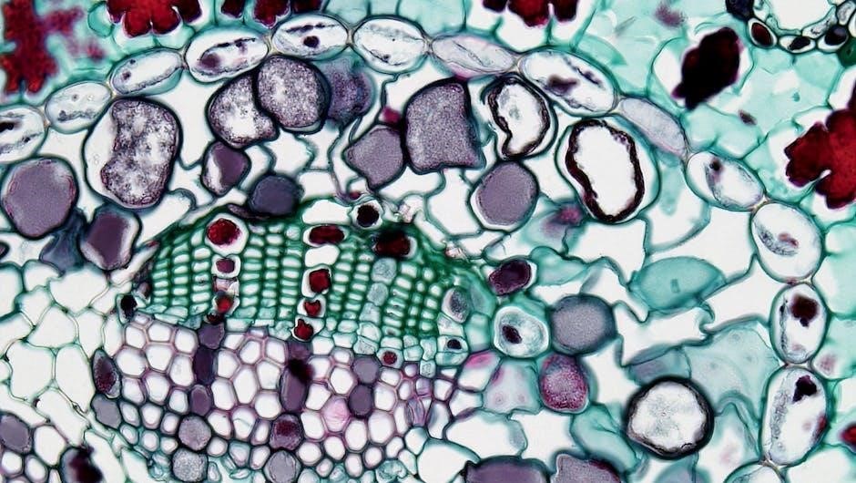
Microscopic Anatomy Coloring
Microscopic anatomy coloring delves into the intricate world of tissues and cells‚ a fundamental aspect of anatomy and physiology. Coloring workbooks dedicated to this level of detail present illustrations of histological structures – epithelial‚ connective‚ muscular‚ and nervous tissues – at a magnified scale.
This process reinforces the identification of cellular components like nuclei‚ cytoplasm‚ and organelles‚ alongside the unique characteristics of each tissue type. Students learn to differentiate between various cell shapes and arrangements‚ crucial for understanding tissue function.
By actively coloring these microscopic views‚ learners solidify their comprehension of cellular structures and their roles within the larger anatomical context‚ building a strong foundation for advanced studies in pathology and physiology.

Choosing the Right Coloring Workbook
Selecting the ideal anatomy and physiology coloring workbook depends on individual learning preferences‚ course requirements‚ and desired level of detail for effective study.
Netter’s Anatomy Coloring Book
Netter’s Anatomy Coloring Book is a highly regarded resource‚ celebrated for its exceptionally detailed and accurate illustrations created by Drs. Frank H. Netter and Carlos Machado.
This workbook features nearly 600 superb illustrations‚ providing essential descriptions of anatomy‚ embryology‚ and pathology‚ making it a comprehensive tool for visual learners.
The book’s strength lies in its ability to reinforce learning through active recall and visual association‚ helping students to identify and remember key anatomical structures.
It’s particularly beneficial for those who appreciate a high level of artistic detail and a thorough exploration of the human body.
Netter’s is available at reasonable prices‚ offering excellent value for students seeking a robust and reliable coloring and study aid.
Princeton Review Anatomy Coloring Workbook
The Princeton Review Anatomy Coloring Workbook is a popular choice‚ offering a comprehensive and accessible approach to learning anatomy and physiology.
It’s designed to simplify the study of these complex subjects through engaging coloring activities and clear‚ concise labeling exercises.
This workbook is particularly useful for students who benefit from a more straightforward and less artistically detailed approach compared to Netter’s.
It provides a solid foundation in anatomical structures and their functions‚ aiding in memorization and comprehension.
Available for free download as a PDF‚ it’s a cost-effective option for students seeking a valuable study resource.
The workbook’s focus on essential descriptions makes it a practical tool for exam preparation and overall understanding.
Other Popular Options
Beyond Netter’s and Princeton Review‚ several other anatomy and physiology coloring workbooks cater to diverse learning preferences. Many students find value in supplementary resources to reinforce their understanding.
Some instructors utilize coloring books in undergraduate courses‚ spanning basic‚ advanced anatomy‚ and even embryology‚ demonstrating their broad applicability.
These alternatives often provide unique perspectives and varying levels of detail‚ allowing students to choose a workbook that aligns with their individual needs.
While specific titles weren’t explicitly mentioned‚ the general consensus highlights the benefit of using coloring as a study aid.
For paramedics‚ EMTs‚ and other healthcare professionals‚ specialized coloring and activity books are available‚ focusing on relevant anatomical structures.
Exploring different options ensures a personalized and effective learning experience‚ enhancing comprehension and retention of key concepts.
Tips for Effective Coloring and Learning
Employing color-coding strategies‚ labeling diagrams‚ and integrating coloring with other study methods maximizes learning and memorization from anatomy workbooks.
Color-Coding Strategies
Color-coding is a powerful technique when utilizing anatomy and physiology coloring workbooks‚ significantly enhancing retention and understanding. Assigning specific colors to different body systems – for example‚ red for cardiovascular‚ blue for respiratory‚ and yellow for nervous – creates visual associations.
This method allows for quick identification and recall of structures and their functions. Consistent color usage throughout the workbook reinforces these connections. Consider using analogous colors for related structures within a system‚ and contrasting colors to differentiate between them.
Furthermore‚ develop a personal color key and consistently refer to it while coloring. This personalized approach caters to individual learning styles and strengthens the link between visual cues and anatomical knowledge‚ ultimately improving study efficiency.
Labeling and Memorization
Labeling diagrams within an anatomy and physiology coloring workbook is crucial for solidifying memorization. Don’t simply color; actively write the names of structures directly onto the illustrations. This kinesthetic learning process – combining visual and tactile elements – dramatically improves recall.
Initially‚ use the workbook’s provided labels as a guide‚ but progressively challenge yourself by recalling names from memory. Flashcards can complement this‚ testing your ability to identify structures without visual cues. Regularly review labeled diagrams‚ spacing out repetitions for long-term retention.
Furthermore‚ create self-tests by covering the labels and attempting to reconstruct them. This active recall strengthens neural pathways‚ making anatomical knowledge more accessible and durable. Consistent labeling reinforces understanding and builds a strong foundation.
Integrating with Other Study Methods
To maximize learning‚ integrate anatomy and physiology coloring with other study techniques. Don’t rely solely on coloring; combine it with textbook readings‚ lecture notes‚ and online resources for a comprehensive approach.
Use the colored diagrams as visual aids during study groups‚ explaining structures and functions to peers. This reinforces your understanding and identifies knowledge gaps. Consider creating mnemonics or stories linking anatomical features to enhance memorization.
Furthermore‚ apply your coloring knowledge to clinical scenarios or case studies‚ bridging the gap between theory and practice. Regularly revisit colored diagrams alongside practice questions‚ solidifying connections and improving exam performance. A multi-faceted approach yields the best results.